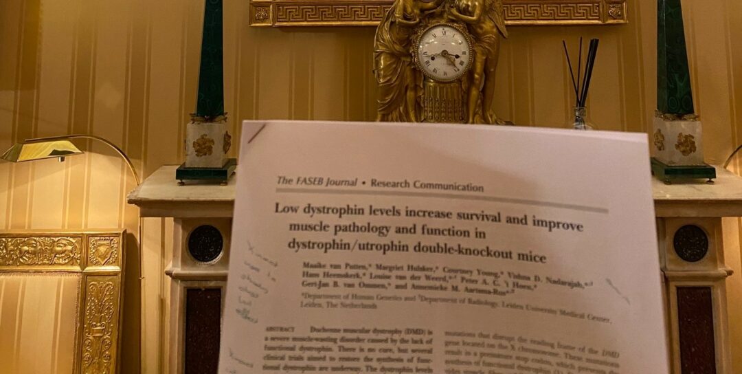
Today’s pick is one of my own papers by van Putten et al in FASEB journal doi 10.1096/fj.12-224170. Here we used the fact that women/females inactivate one of their X-chromosomes to study how much dystrophin is needed for survival and function.
Low dystrophin levels increase survival and improve muscle pathology and function in dystrophin/utrophin double-knockout mice
X-inactivation also occurs in mice, normally in the 50:50 random pattern. However, there is a mouse model where the gene (Xist) coordinating X-inactivation has been altered. The chromosome with this altered Xist is more likely to be inactivated. This means that if you cross females with this Xist mutation with mdx males, the resulting female offspring will preferentially inactivate the X-chromosome with the Xist mutation and a functional dystrophin gene.
As the process is skewed but still random, this results in females expressing anywhere between 2 and 40% dystrophin in their muscles. We first did this in mdx mice, but they are only mildly affected. Here, we studied this in an mdx/utrophin knockout (dko) background, so we could also study impacts on survival.
In a first study we subjected mice to a 12 week functional test regime starting at 4 weeks of age. DKO mice (no dystrophin) generally had to be humanely killed before the age of 12 weeks. The dko/xist mice (having between 3-16% dystrophin in their muscle) survived longer. We subdivided the mice in those having <4% dystrophin and those having >4% dystrophin.
Even mice with <4% dystrophin had increased survival, lower CK and better function than dko, however mice with >4% dystrophin performed even better – though still less than wild type. We then performed a long term survival study. Notably in these studies we could only see the dystrophin levels after the animals had been sacrificed (a blinded experiment).
The 6 dko mice all died before 12 weeks, while those with <4% dystrophin survived longer. For those with 4-15% dystrophin 64% was alive at 10 months, while all mice with >15% were still alive at this point. These mice also resembled wild types in function and appearance.
6 mice were randomly selected for a very long survival study. The mice with 3% and 27% dystrophin died at 17 and 25 months due to muscular dystrophy. Other mice (>30% dystrophin) died due to age related causes >2 years old or a gall bladder problem (17 months).
Additional studies showed that dystrophin levels correlated with TIMP-1 serum levels (in mouse – we have not been able to translate this finding for patients). Also when looking at muscle histology we observed improvements at <4% dystrophin but more improvement at higher levels.
While this now may seem like old news (the paper is from 2013), it was one of the first papers showing that little bits of dystrophin already can go a long way and that it was likely not required to restore dystrophin at 20% to have a functional effect (as was the consensus at that time)
There are 3 limitations to this study however 1. The study had to use females, while Duchenne primarily occurs in men. 2. The mice had dystrophin from birth, while in patients restoration of dystrophin occurs later in life. 3. The study was in mice and not in human, so the levels may not translate directly.


